Article Text

Abstract
Background There have been no effective treatments for slowing or reversing Alzheimer’s disease (AD) until now. Growing preclinical evidence, including this study, suggests that mesenchymal stem cells-secreted exosomes (MSCs-Exos) have the potential to cure AD.
Aims The first three-arm, drug-intervention, phase I/II clinical trial was conducted to explore the safety and efficacy of allogenic human adipose MSCs-Exos (ahaMSCs-Exos) in patients with mild to moderate AD.
Methods The eligible subjects were assigned to one of three dosage groups, intranasally administrated with ahaMSCs-Exos two times per week for 12 weeks, and underwent follow-up visits at weeks 16, 24, 36 and 48.
Results No adverse events were reported. In the medium-dose arm, Alzheimer’s Disease Assessment Scale–Cognitive section (ADAS-cog) scores decreased by 2.33 (1.19) and the basic version of Montreal Cognitive Assessment scores increased by 2.38 (0.58) at week 12 compared with baseline levels, indicating improved cognitive function. Moreover, the ADAS-cog scores in the medium-dose arm decreased continuously by 3.98 points until week 36. There were no significant differences in altered amyloid or tau deposition among the three arms, but hippocampal volume shrank less in the medium-dose arm to some extent.
Conclusions Intranasal administration of ahaMSCs-Exos was safe and well tolerated, and a dose of at least 4×10⁸ particles could be selected for further clinical trials.
Trial registration number NCT04388982.
- Neurocognitive Disorders
Data availability statement
All data relevant to the study are included in the article or uploaded as supplemental information.
This is an open access article distributed in accordance with the Creative Commons Attribution Non Commercial (CC BY-NC 4.0) license, which permits others to distribute, remix, adapt, build upon this work non-commercially, and license their derivative works on different terms, provided the original work is properly cited, appropriate credit is given, any changes made indicated, and the use is non-commercial. See: http://creativecommons.org/licenses/by-nc/4.0/.
Statistics from Altmetric.com
WHAT IS ALREADY KNOWN ON THIS TOPIC
Mesenchymal stem cells-secreted exosomes (MSCs-Exos) can alleviate Alzheimer’s disease (AD) in mouse models. However, there was no evidence suggesting whether MSCs-Exos are suitable for use in AD clinical treatment.
WHAT THIS STUDY ADDS
This study conducted the first clinical trial of MSCs-Exos in patients with AD and demonstrated their safety in humans.
HOW THIS STUDY MIGHT AFFECT RESEARCH, PRACTICE OR POLICY
This study provided dose selection for further phase II and phase III clinical trials. MSCs-Exos could provide hope for the treatment of AD if proven effective in multicentred, double-blinded, randomised controlled trials.
Introduction
Alzheimer’s disease (AD), the most common neurodegenerative disease, is the leading cause of dementia and is the fifth leading cause of death in the elderly.1 Although numerous attempts have been made to develop drugs for AD, there are currently no effective treatments for halting or reversing the progress of AD.2 The development of novel clinical available drug pipelines for the treatment of AD is therefore required.
In recent years, exosomes have been found to play an important role in AD,3 and may provide a novel diagnostic or therapeutic approach for AD management. Exosomes are extracellular vesicles that are released by diverse cells in rest or stress states and contain enhanced lipids, proteins and nucleic acids. They can be delivered to distant regions via bodily fluids, and taken up by target cells via three pathways: membrane fusion, endocytosis and the ligand receptor-mediated mechanism.4 Mesenchymal stem cells (MSCs)-derived exosomes provide unparalleled advantages over MSCs, including low immunogenicity, bulk synthesis from commercially available cell lines, avoidance of invasive MSCs in collection or differentiation, and simplicity in storage.5
Due to their capacity to reach the brain, exosomes released by MSCs shed new light on developing disease-modifying strategies for AD.6 Previous limited investigations demonstrated that intracerebroventricular injections7 and the systematic injection8 of MSCs-derived exosomes ameliorated cognitive impairment in AD animals. In addition, our team has established the potential of allogenic human adipose-derived MSCs exosomes (ahaMSC-Exos) from healthy volunteers for the intranasal treatment of AD mice. After intranasal administration with a dose of 30 µg ahaMSCs-Exos per mouse, ahaMSCs-Exos can be rapidly transported to various brain regions through the olfactory system involving olfactory sensory neurons. ahaMSCs-Exos in the brain reached a peak at 1 hour after administration and were mainly taken up by neurons. In APP/PS1 mice, ahaMSCs-Exos displayed neuroprotective effects, increased neurogenesis and enhanced spatial memory. ahaMSCs-Exos also appeared to decrease amyloid beta deposition and inhibit microglial activation. Importantly, ahaMSCs-Exos did not induce morphological alterations in key organs, indicating the safety of the intranasal administration.9 Another study demonstrated that an intranasal injection of MSCs-derived exosomes might improve AD by suppressing microglia cell activation and boosting dendritic spine density in triple-transgenic 3xTg mouse models.10 These findings clearly demonstrate the therapeutic potential of MSCs-derived exosomes for AD and call for testing in real clinical settings. Inspiringly, in preclinical settings, MSCs-derived exosomes have been used to treat inflammatory and degenerative disorders, such as lung damage diseases, wound healing, liver diseases, cardiovascular diseases and bone regeneration.11 Moreover, there is growing evidence that MSCs-derived exosomes can be applied for the management of neurological diseases, such as ischaemic stroke,12 neurodegenerative diseases,13 spinal cord injury,14 etc. However, MSCs-derived exosomes have not been clinically investigated in AD despite the disease-modifying effect they have on transgenic mice models.
Accordingly, here, we conducted the first clinical trial to investigate the safety and efficacy of intranasal administration of ahaMSCs-Exos in patients with AD, aiming to demonstrate its application value in clinical settings and provide a solid foundation for future large-scale multicentre studies.
Methods
Exosomes derived from allogenic adipose MSCs
The intranasal drug (ahaMSCs-Exos) was produced by Cellular Biomedicine Group (Shanghai, China, https://www.cellbio.com) according to the previously reported manufacturing and quality control process.15 Specifically, after liposuction, healthy young adult donors’ adipose tissue was used to separate fat cells for the creation of ahaMSCs. All cell-related operations were performed in a Current Good Manufacturing Practice (cGMP)-compliant laboratory. The manufacture was optimised to combine precipitation and ultracentrifugation based on previous studies with polyethylene glycol applied for the enrichment of ahaMSCs-Exos.16 17 The production process achieved 3×10¹⁰–5×10¹⁰ nanoparticles from 500 mL condition medium. Quality control standards were established according to the process development and regulations of cell-based products: (1) the morphology of the product is a cup-shaped vesicle with a double membrane, which was observed by transmission electron microscopy after staining with 3% uranyl acetate, according to our previous method9 (online supplemental figure 1A); (2) more than 80% of the particles are between 30 and 160 nm, which is compatible with the exosome diameter (30–150 nm), and the size distribution was analysed by nanoparticle tracking analysis (NanoSight NS300, Malvern) (online supplemental figure 1B); (3) marker expression includes CD63+, CD81+, CD9+, TSG101+ and CANX− (online supplemental figure 1C), which were measured by western blot; (4) other requirements for cell-based products, such as sterility (negative), endotoxin (< 100 EU/mL) and Mycoplasma (negative).
Supplemental material
Study design of the open-label, phase I–II clinical trial
The study was registered (ClinicalTrials.gov NCT04388982) and approved by the Ethics Committee. It is a three-arm, open-label, single-centre, drug-intervention, phase I/II clinical trial, aiming to estimate the safety and explore the efficacy of ahaMSCs-Exos in the treatment of AD. The trial, which took place from April 2020 to April 2022, was composed of three periods for each participant: the screening period (21 days), the intervention period (12 weeks) and the follow-up period (48 weeks). After enrolment, each participant was assigned the sequence number according to the time of inclusion. During the intervention period, each participant undertook an intranasal administration of ahaMSCs-Exos (2×10⁸, 4×10⁸, 8×10⁸ particles) diluted in saline (1 mL) two times per week (24 treatments in total) through nasal spray devices. Simultaneously, the 3+3 design was used in the three groups divided according to the interventional dose from low to high. For instance, if no case of dose-limited toxicity (DLT) existed in the three subjects of the low-dose group, the clinical trial moved to the middle-dose group. If one case of DLT existed, an additional three subjects were added, with all subjects accepting medical observation. Theoretically, 3–18 eligible subjects were required. The flow of this trial is depicted in figure 1.

Study design of the clinical trial. n=3−18 (theoretically). DLT, dose-limited toxicity.
Procedures
The ahaMSCs-Exos were shipped in a frozen state to the clinical site in a dry ice container with a continuous temperature monitor. Upon receipt, the solution was examined and stored in a regulated, −80°C storage tank that was regularly monitored. All transfers and the signing paperwork were documented. Before nasal spray administration, the solutions were thawed at room temperature and shaken. Each batch of the drug was valid for 30 days.
At baseline, basic information, medical history and vital signs were recorded. Laboratory tests were also conducted, including a 12-lead electrocardiogram (ECG). Laboratory tests included the following items: infectious diseases tests, blood routine, urinalysis, hepatic function, renal function, fasting plasma glucose, blood lipids, coagulation function and immunology. During the treatment period, the melted drug was shaken to mix and administered alternately through the left and right nasal cavities, where it was sprayed about once a minute for 10 min. The time of the beginning and ending was recorded, as well as vital signs, symptoms of adverse effects (AEs) or serious AEs (SAEs). Laboratory tests (excluding tests for infectious diseases) and ECG were performed at the end of the 8th treatment (4 weeks), 24th treatment (12 weeks) and at the first follow-up visit (16 weeks). All participants accepted cognitive examinations and an evaluation for quality of life at the baseline, after 24 treatments (12 weeks) and during the follow-up period (16 weeks, 24 weeks, 36 weeks, 48 weeks) (figure 2).

Flowchart and specific visits for each participant. ECG, electrocardiogram;PET, positron emission tomography.
Participants
All participants were enrolled from the Memory Clinic of Neurology Department and informed consent was obtained. Each participant should meet all items of the following criteria: (1) ≥50 years; (2) diagnosed with mild or moderate AD, according to the recommendations from the National Institute on Aging and Alzheimer’s Association 201118 and the updated research framework in 201819; (3) suspension of cognitive-enhancing drugs and marketed therapeutic drugs such as ginkgo, high-dose of vitamin E, lecithin, oestrogen, non-steroidal anti-inflammatory drugs, donepezil, memantine, and so on.
Subjects were excluded if any of the following conditions were present: (1) suffering from severe and poorly controlled concomitant diseases or with severe allergic constitution; (2) diagnosis of severe depression or other psychiatric disorders or neurological diseases; (3) MRI contraindications; (4) participation in any other clinical trial within 6 months. Detailed inclusion and exclusion criteria were listed in online supplemental materials.
Outcome measures
The safety of the ahaMSCs-Exos administration was assessed at baseline, and 4, 12, 16 weeks after the first treatment, based on vital signs, clinical laboratory tests, AEs and SAEs.
The efficacy evaluation consisted of assessments of cognitive function and daily activities. The outcome measures included scores according to the Alzheimer’s Disease Assessment Scale–Cognitive section (ADAS-cog, scores range from 0 to 70),20 the Mini-Mental State Examination (MMSE, scores range from 0 to 30),21 the basic version of Montreal Cognitive Assessment (MoCA-B, scores range from 0 to 30),22 the Neuropsychiatric Inventory (NPI, scores range from 0 to 144 for patients and from 0 to 60 for caregivers, respectively),23 the scale of Alzheimer’s Disease Cooperative Study–Activities of Daily Living (ADCS-ADL),24 the Activities of Daily Living Scale (ADL) and Instrumental Activities of Daily Living Scale (IADL) at baseline, and 12, 16, 24, 36 and 48 weeks after the first treatment. As the higher score of ADAS-cog stands for a worse cognitive level, declined ADAS-cog scores indicate the cognitive improvement. On the contrary, scores of MMSE or an MoCA-B increase represent an improvement in cognition.
The primary endpoint was defined as (1) the number of participants with treatment-related abnormal laboratory values of hepatic or kidney function, and (2) the number of participants with treatment-related AE/SAE as assessed by the Common Terminology Criteria for Adverse Events-V.4.0. An interim analysis was performed during the trial.
Positron emission tomography and hallmarks of AD pathology
Positron emission tomography (PET) imaging was used to evaluate the hallmarks of AD pathology. The PET-MRI for amyloid and the PET-CT for tau were conducted at the baseline and the last visit of the follow-up period. Participants were injected with 18F-florbetapir intravenously at a dose of 5 mL/kg and underwent PET scanning which lasted 20 min at 1 hour after injection. Standard uptake value ratios (SUVRs) were calculated to quantify the cerebral amyloid plaques and tau deposition. Regions of interest (ROIs) included frontal cortex, parietal cortex, lateral temporal, precuneus (PreC), anterior cingulate cortex and posterior cingulate cortex (PCC) for amyloid, whereas lateral parietal, lateral temporal, medial temporal, frontal, occipital cortex, PreC and PCC for tau. A similar procedure was operated in tau imaging which employed 18F-PM-PBB3 as the PET tracer. Hippocampal volumes were also calculated based on the T1-weighted MRI.
Statistical analysis
Considering the small sample size, descriptive statistics were used. The mean (standard deviation, SD) was described for the measurement data. The changes in scores of scales from baseline to completion of treatments (12 weeks) were analysed by a paired t-test. Analysis of variance was adopted in the comparison of the following means of the three arms. For hallmarks of AD pathology, the percentage of alterations in amyloid/tau deposition and hippocampal volumes was calculated through (changes/baseline value)×100%. To compare the difference in alterations in amyloid and tau deposition, the percentage of altered SUVRs (%) of different cerebral regions was put together and compared among the three arms. The rates of change in volumes of the bilateral hippocampus were also compared among the three arms. SPSS V.22.0 and GraphPad Prism V.9.0 were employed in the statistical analysis. A p value of <0.05 was considered significant.
Results
Demographical information
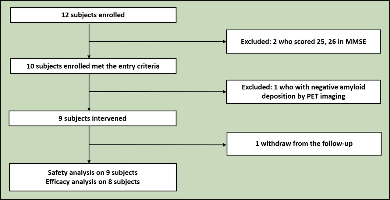
After enrolment, a total of nine eligible participants (figure 3), five males and four females, were intranasally administrated with ahaMSCs-Exos (table 1). Among them, five subjects were diagnosed with mild AD at the baseline, while the other four were diagnosed with moderate AD. They were sequentially assigned to three different treatment arms with 2×10⁸ particles within 1 mL solution for the low-dose arm, twofold for the medium-dose arm and fourfold for the high-dose arm. Demographical information can be found in table 1. The first subject (E001) undertook intranasal administration of ahaMSCs-Exos from 18 July 2020 to 7 October 2020, while the last subject (E009) from 15 May 2021 to 10 August 2021. All subjects completed the treatments, one subject (E003) missed one follow-up appointment, while another (E009) dropped out of the follow-up owing to personal reasons.

The inclusion, exclusion and withdrawal of the enrolled participants. MMSE, Mini-Mental State Examination; PET, positron emission tomography.
The demographic information of the subjects who received the intervention of ahaMSCs-Exos at baseline.
Safety of intranasal ahaMSCs-Exos administration
No AEs or SAEs were reported during the treatment period, nor did any subjects report nasal discomfort during treatment (online supplemental table 1). Blood pressures (figure 4A) and heart rates (figure 4B) fluctuated without obvious abnormalities and a single subject (E009) had occasional bradycardia. No allergic events occurred during the intervention in all subjects despite the high level of IgE in subjects E001 and E007 (figure 4C). Laboratory tests did not suggest that the subjects had treatment-related damage in hepatic or kidney function. As depicted in figure 4D–G, the concentrations of alanine transaminase, aspartate aminotransferase, blood urea nitrogen and serum creatinine remained stable and within normal ranges.

Safety evaluations of the intranasal ahaMSCs-Exos administration. (A) Blood pressure of each subject at baseline and 24 interventions (systolic and diastolic blood pressures); (B) heart rates of each subject at baseline and 24 interventions with a normal range of 60–100 beats per minute (bpm); (C) IgE levels; (D,E) laboratory indicators for liver function: alanine transaminase (ALT) and aspartate aminotransferase (AST); (F,G) laboratory indicators for renal function: blood urea nitrogen (BUN) and serum creatinine (Scr). (C–G) The indicators level at baseline, during the intervention period and 4 weeks after intervention. ahaMSCs-Exos, allogenic human adipose mesenchymal stem cells-secreted exosomes.
Efficacy of intranasal ahaMSCs-Exos administration: from baseline to the completion of treatment periods (12 weeks)
Changes in the scores of ADAS-cog (1.4 (5.4), p=0.500) and MoCA-B (−1.1 (4.6), p=0.510) at 12 weeks were not significant compared with the baseline (online supplemental table 2), while scores of MMSE remained unchanged (0.0 (4.0), p=1.000). Moreover, the neuropsychiatric symptoms and the daily living abilities did not differ significantly after administration. Caregivers’ worries about the patients’ neuropsychiatric symptoms seemed to be reduced (−2.4 (3.7), p=0.136, did not reach statistical significance) (online supplemental table 2).
To explore whether an interventional dose had an influence, a stratified analysis was performed. Results diverged between the different dose arms. Changes in scores of cognitive scales were inconsistent in the subjects administrated with a low dose, and also in the high-dose arm (figure 4). However, ADAS-cog scores showed a consistent trend in the medium-dose arm, decreasing by 2.33 (1.19) compared with the baseline (figure 5A). Scores of MMSE had few increases in the high-dose arm (figure 5B). Similarly, an improved trend was observed in the scores of MoCA-B: the MoCA-B scores increased by 2.38 (0.58) in the medium-dose arm compared with the baseline, suggesting a better cognitive performance (figure 5C). In addition, the caregivers of the subjects who received the medium-dose intervention had a reduction of 5.33 (3.22) in distress due to neuropsychiatric symptoms based on the NPI. However, the neuropsychiatric symptoms of the patients did not ameliorate, in fact, they worsened (figure 5D). There were no significant discrepancies in changes in scores of depression observed among the three arms (figure 5E). Furthermore, in the medium-dose arm, improvements in activities of daily living were indicated by an increasing trend in scores of ADCS-ADL and a decreasing trend in scores of ADL to some extent (figure 5F).

Changes in scores of scales from baseline to week 12. (A–C) Cognition examination scales: Alzheimer’s Disease Assessment Scale–Cognitive section (ADAS-cog), the Mini-Mental State Examination (MMSE) and the basic version of Montreal Cognitive Assessment (MoCA-B). (D) The Neuropsychiatric Inventory Scale (NPI). (E) The Geriatric Depression Scale (GDS). (F) Assessments of daily living activities: the scale of Alzheimer’s Disease Cooperative Study–Activities of Daily Living (ADCS-ADL), the Activities of Daily Living Scale (ADL) and the Instrumental Activities of Daily Living Scale (IADL).
Based on the results, the intranasal administration of ahaMSCs-Exos might be effective in the medium-dose arm (at a dose of 4×10⁸ particles, 1 mL).
Changes in the scores of the neuropsychological scales in the follow-up period
After the 24 intranasal administrations (12 weeks), the scales were performed on the participants at weeks 16, 24, 36 and 48, respectively. ADAS-cog scores were in continuous decline by 3.98 in the medium-dose arm until week 36, while the MMSE and MoCA scores remained relatively stable in the follow-up period (online supplemental figure 2A–C). It could therefore be inferred that the ahaMSCs-Exos possess post-effects to maintain cognition for an extra 6 months after the treatment, lasting about 3 months. MMSE and MoCA scores indicated that cognitive function worsened in the low-dose and high-dose arms, despite the resumption of usual medication following intervention or in follow-up. The scores of NPI and Geriatric Depression Scale fluctuated without apparent tendencies, suggesting that neuropsychiatric symptoms and depression states fluctuated throughout this trial among all the subjects (online supplemental figure 2D–F). The trend of changes in the ADL scores was consistent with that of the cognitive assessments to some extent. Combined scores of ADCS-ADL, ADL and IADL scales and daily living abilities could allow an additional 6 months to be maintained in the medium-dose arm (online supplemental figure 2G–I).
Changes to AD pathological hallmarks in the follow-up period
Based on the ATN (amyloid, tau, neurodegeneration) profile of AD pathological hallmarks, for A, data from PET-MRI suggest that the cerebral amyloid plaques increased approximately 1 year following the first treatment (online supplemental table 3). The exception was the declined SUVRs of several regions observed in the subject E007 with the average of ROIs from 1.09 to 1.01, indicating reduced cerebral amyloid deposition in this participant, particularly in the PreC. There were no significant differences among the three-dose arms from the perspective of proportions of altered amyloid deposition compared with the baseline (figure 6A). For T, the level of tau deposition showed no obvious change trend a year after follow-up (figure 6B). For N, in the medium-dose arm, the volume of the left hippocampus decreased by 0.118 (0.068), and 0.213 (0.289) for the low-dose and 0.181 (0.037) for high-dose groups, respectively. Similarly, the volume of the right hippocampus reduced: 0.290 (0.325), 0.076 (0.061) and 0.161 (0.072) in three arms. Hippocampal volume shrank less in the medium-dose arm but did not reach statistical significance as the rate of changes in hippocampal volumes did (figure 6C).

Changes of hallmarks of AD pathology (amyloid, tau, neurodegeneration) in the follow-up period compared with baseline. The violin plot of the amyloid deposition (A) and tau deposition (B) alterations after 1 year detected by PET-MRI, comparing with baseline. The selected regions of interest included the frontal cortex, parietal cortex, lateral temporal, precuneus, anterior cingulate cortex and posterior cingulate cortex for amyloid, whereas lateral parietal, lateral temporal, medial temporal, frontal, occipital cortex, precuneus and posterior cingulate cortex for tau. (C) Altered proportions of bilateral hippocampal volumes indicated less atrophy in the subjects who accepted medium-dose administration. AD, Alzheimer’s disease; MRI, magnetic resonance imaging; PET, positron emission tomography.
Discussion
Main findings
To the best of our knowledge, this trial is the first attempt to apply MSCs-secreted exosomes for the clinical treatment of AD. The results demonstrated the safety of an intranasal administration of ahaMSCs-Exos for AD treatment, and a dose of at least 4×10⁸ particles could be selected for further large-scale multicentre clinical trials. First, no mild AEs or severe AEs were reported during the treatment period. Subjects with high IgE levels at baseline were also tolerant to the nasal administration. Second, although for all the subjects changes in the scores of cognitive scales at week 12 were not significant compared with the baseline, in the medium-dose group, ADAS-cog scores consistently decreased by 2.33 (1.19) and MoCA-B scores increased by 2.38 (0.58) at week 12 compared with the baseline, indicating improved cognitive function. Lastly, we revealed the microRNA (miRNA) and protein expression profiles in ahaMSCs-Exos, which identified 277 miRNAs and 1443 proteins, and supplied the potential neurotrophic factors for patients with AD in the current clinical trial.
In terms of the present pilot trial, ahaMSCs-Exos at a dose of 4×10⁸ particles once (the medium dose) had potential for the treatment of AD. The cognitive function of the subjects administrated with medium-dose ahaMSCs-Exos improved after the intervention, as reflected by decreased ADAS-cog scores and increased MoCA-B scores. Additionally, the subjects in the medium-dose arm had better performance in daily living activities compared with the baseline. The amelioration could be maintained for an extra 6 months following the 3-month treatment period due to a possible after-effect. Treatment effects may be attributed to slowing hippocampal atrophy to some extent. Exosomes carry and transfer a variety of functional molecules such as proteins, DNAs, lipids and RNA with regulatory effects, which can modify cell metabolism. In the proteomic characterisation of the MSC-exosomes, hundreds of proteins have been identified, some of which are specific cell type markers and others are involved in the regulation of the binding and fusing of exosomes with recipient cells. Some factors promoting the recruitment, proliferation and differentiation of neural stem cells and other cells were also found. Moreover, a wide range of miRNAs have been found in exosomes, which can control functions related to nerve remodelling, angiogenesis and neurogenesis.25 26 Taking this into consideration, the use of exosomes may be part of a strategy to promote neural plasticity in AD, improving cognitive impairment and neural replacement.
Considering that the two subjects in the high-dose arm had high ADAS-cog scores at the baseline, which indicated relatively poor cognitive status before administration, it was inferred that the ahaMSCs-Exos might be insensitive to patients with moderate to severe AD, at least in terms of the 8×10⁸ particle dose. Despite no cognitive improvement, a reduction in the amyloid plaque burden was observed in a subject in the high-dose arm (E007), which deserved in-depth study. Furthermore, neither of the two patients with early-onset disease experienced cognitive improvement after treatment. Therefore, further studies should focus on patients with mild AD and those with mild cognitive impairment due to AD. The patient’s age at onset should also be taken into consideration. It is difficult to elucidate whether higher doses are effective and the optimal frequency of treatment, so both need further research.
The therapy effect of ahaMSCs-Exos may be equivalent or better when compared with drugs for AD on the current market. The ADAS-cog scores changed (−2.33 at week 12, –3.21 at week 16, –3.52 at week 24 and −3.98 at week 36) in patients given a medium dose of ahaMSCs-Exos. The published results suggested that the ADAS-cog scores declined (2.58 (5.7)) in the 900 mg group following the 24-week treatment (p=0.30 in comparison with the placebo group) for the sodium oligomannate (GV-971).27 The phase III clinical trial of GV-971 demonstrated that the ADAS-cog scores changed (−1.89 (95% confidence interval (CI): −2.78 to −1.00)) at week 36 in the drug group and the difference was −2.15 (95% CI: −3.07 to −1.23) (p<0.001) between GV-971 and the placebo.28 For donepezil, the differences between the ADAS-cog scores were −2.1 in the 5 mg group and −2.7 in the 10 mg group at week 12 compared with the baseline.29
Furthermore, both miRNAs and proteins in ahaMSCs-Exos were analysed through RNA sequencing and proteomic analysis. It was suggested that the benefits of ahaMSCs-Exos could be due to supplementing miRNAs which are reduced in patients with AD and proteins that regulate the exosome-related and neuroimmune-related signal pathways (online supplemental tables 4 and 5 and figures 3 and 4). However, the relatively specific mechanism of ahaMSCs-Exos on AD needs further elucidation.
Limitations
Limitations existed in this trial. On the one hand, the study is a pilot clinical trial with a relatively small sample size for undertaking a statistical comparison; on the other hand, the second PET scan was conducted 1 year after the baseline, meaning it was challenging to determine whether amyloid or tau reduced after administration, as AD had progressed. Some confounding effects existed based on the current inclusion criteria, such as the age at onset, dementia stage, common comorbidities, and so on. In further clinical trials, stricter inclusion criteria will be set to clarify the effectiveness of ahaMSCs-Exos. The traditional 3+3 design with dose increments of 100% was adopted in this trial. Based on the results, the safety of ahaMSCs-Exos through nasal administration was proven and there seemed to be no delay or cumulative toxicity at experimental doses. The maximum tolerated dose (MTD) was not determined due to the relatively conservative dosing design. In further trials, accelerated titration design or pharmacologically guided dose escalation could be applied to determine the MTD. Additionally, selecting bias, measurement bias and withdrawal bias were included in this trial.
Implications
The intranasal administration of ahaMSCs-Exos was safe and well tolerated at a treatment frequency of two times per week. The dose of at least 4×10⁸ particles could be selected for a randomised phase II and phase III clinical trial in further steps. Improvements should be performed in next-phase clinical trials, such as expanding the sample size, setting the control group and stricter inclusion criteria, extending the intervention period, and adding trajectories of imaging and biomarkers of peripheral blood.
Data availability statement
All data relevant to the study are included in the article or uploaded as supplemental information.
Ethics statements
Patient consent for publication
Ethics approval
This study involves human participants and was approved by the Ethics Committee of Ruijin Hospital, Shanghai Jiao Tong University School of Medicine (2019-249). Participants gave informed consent to participate in the study before taking part.
Acknowledgments
Sincere thanks to each subject participating in this clinical trial. We thank Li Xia, Shuang Meng and Qin Fu from the Core Facility of Basic Medical Sciences, Shanghai Jiao Tong University School of Medicine for the analysis of proteomics. We express our sincere appreciation to the following individuals for their support and assistance for ahaMSCs-Exos production: Jing Wang, Jigang Lei, Cuili Xu, Meiping Shen, Zeyi Zhang, Chengjie Ren and Bizuo Liu from the Cellular Biomedicine Group.
References
Xinyi Xie graduated from Tongji Medical School, Huazhong University of Science and Technology, China in 2018 and obtained her master’s degree in neurology at Ruijin Hospital, Shanghai Jiao Tong University School of Medicine, China in 2021. Dr Xie is currently working as a resident at the Department of Neurology, Shanghai Ninth People's Hospital, affiliated to Shanghai Jiao Tong University School of Medicine, China. During the master’s period, under the supervision of Prof. Rujing Ren and Prof. Gang Wang, the research field focused on neurodegenerative diseases, especially Alzheimer’s Disease and related cognitive impairments. The main research results were published on Genes & Diseases, JAD and so on.

Supplementary materials
Supplementary Data
This web only file has been produced by the BMJ Publishing Group from an electronic file supplied by the author(s) and has not been edited for content.
Footnotes
XX, QS, CD and SC contributed equally.
Contributors Conceptualisation—GW, XG and RR. Methodology—XX, QS, RR, CD, RT, SC, SL, JC, PL, XG and GW. Investigation—XX, QS, RR, RT, SC, JW, JL, CG, SDC, XG and GW. Visualisation—XX, QS, RR, RT, XG and GW. Funding acquisition—GW, XG and QS. Project administration—SDC, XG and GW. Supervision—GW, XG, SDC and HC. Writing (original draft)—XX, QS, SC and RT. Writing (review and editing)—GW and XG. Guarantor—GW.
Funding This work was supported by the Ministry of Science and Technology of the People's Republic of China (2021ZD0201804, GW); National Natural Science Foundation of China (92068111, 81973272, XG; 81903582, QS); Natural Science Foundation of Shanghai (219ZR1431500, GW); Shanghai Science and Technology Committee (121XD1422200, XG) and Cellular Biomedicine Group (CBMG, Shanghai, China).
Competing interests CD, SL, JC and PL are current employees of Cellular Biomedicine Group. Other authors declare that they have no competing interests.
Provenance and peer review Not commissioned; externally peer reviewed.
Supplemental material This content has been supplied by the author(s). It has not been vetted by BMJ Publishing Group Limited (BMJ) and may not have been peer-reviewed. Any opinions or recommendations discussed are solely those of the author(s) and are not endorsed by BMJ. BMJ disclaims all liability and responsibility arising from any reliance placed on the content. Where the content includes any translated material, BMJ does not warrant the accuracy and reliability of the translations (including but not limited to local regulations, clinical guidelines, terminology, drug names and drug dosages), and is not responsible for any error and/or omissions arising from translation and adaptation or otherwise.